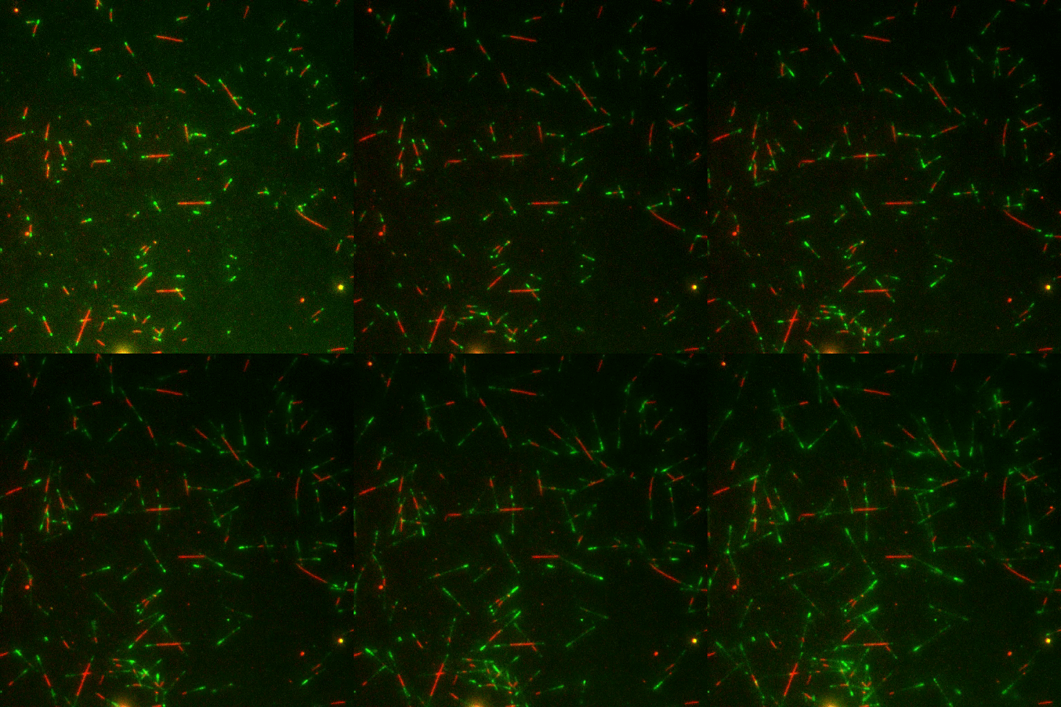
In vitro TIRF microscopy
Direct measurement of MAP and Microtubule interaction
Cells are messy. In order to definitively determine the influence of Microtubule-Associated Proteins (MAPs) on microtubules, we assay single microtubules in the presence of purified MAPs under controlled biochemical conditions.

Electron Microscopy
Structural Biology using Cryo-EM
The complex of microtubules and MAPs cannot be crystallized. Cryo-EM enables us to solve protein structures like MAPs bound to microtubules at near-atomic resolutions from a lot of really blurry images. In addition we can directly visualize the structure of the microtubue lattice and end.

Cell Biology
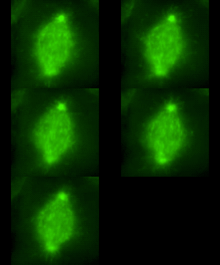
Manipulation and Imaging of MAPs and Microtubules in Cells
Our ultimate goal is to understand what MAPs in cells are doing and how we can exploit that knowledge for the development of new drug targets. We are especially interested in MAPs that help building the mitotic spindle. A fine tuned machinery of dozens of different MAPs help to make and break microtubules that are building the spindle. Abbereations in spindle assembly often result in errors in chromosome segregation. The resulting chromosomal instability is a hallmark of human cancer and can drive both the initiation and progression of tumors. We are assaying for MAP localization and phenotypes using CRISPR/Cas9 and optogenetic techniques.
